Dental X-ray examination (dental X-ray) is the basis for quality treatment with a good prognosis. Without high-quality X-ray diagnosis it is almost impossible to make a competent treatment plan. Dentists use several types of X-ray examinations, which allow to examine both one tooth separately and the whole jaw: bone structures, joints, connective tissues. X-ray examination is the basis for quality treatment with a good prognosis. Without high-quality X-ray diagnosis it is almost impossible to make a competent treatment plan. Dentists use several types of X-ray examinations, which allow to study both one tooth separately and the whole jaw: bone structures, joints, connective tissues.
- Intra-oral radiographic techniques is the most common type. Performed on a visiograph with low radiation exposure. This method is used diagnose in the area of 1-2 teeth.
- Panoramic radiography , also called panoramic x-ray, is a two-dimensional (2-D) dental x-ray examination that captures the entire mouth in a single image
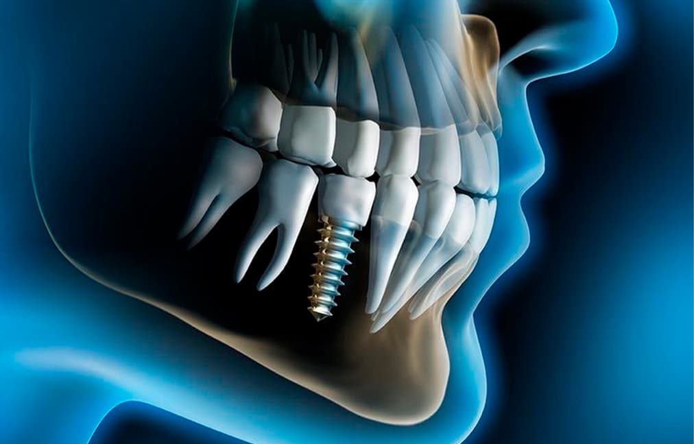
- Dental computed tomography (3D image). Today this is the standard in dentistry. CT is required for volumetric therapeutic treatment, in preparation for implantation, dentures, orthodontics. With its help, it is possible to identify deep lesions of the teeth, bone changes, and malocclusion.
X-rays of the teeth are necessary when carrying out therapeutic treatment (especially when treating canals), prosthetics, implantation, as well as before starting orthodontic treatment (bite correction).
- inflammation inside the tooth;
- presence, size and location of the cyst;
- presence of a tumor;
- quality of dental canal filling;
- deeps of caries ;
- abscess in the periodontal elements;
- crack in the tooth;
- the presence of uncut teeth (including wisdom teeth);
- various anomalies in the structure of bones;
Dental X-rays helps to detect:
What is an orthopantomogram for?
In order to see not only the teeth to be treated or dentured, but also the overall ratio of roots in the dentition. In addition, a panoramic image of the teeth shows not only the roots of the teeth, but it also shows the surrounding periodontal tissues. With its help the doctor sees the location of the roots of the teeth relative to the nerves (mandibular, maxillary) and maxillary sinuses. This allows to determine the optimal type of treatment and avoid any surprises and side effects.
In many cases, the doctor needs to see not only the teeth to be treated or prosthetics, but also the overall ratio of roots in the dentition. In addition, the panoramic image allows to evaluate the amount of bone tissue and their structure and provides a lot of other valuable information. Finally, orthopantomography reveals hidden pathologies — inflammation, cavities, cysts, periodontal pockets, makes it possible to take timely measures to prevent complications.
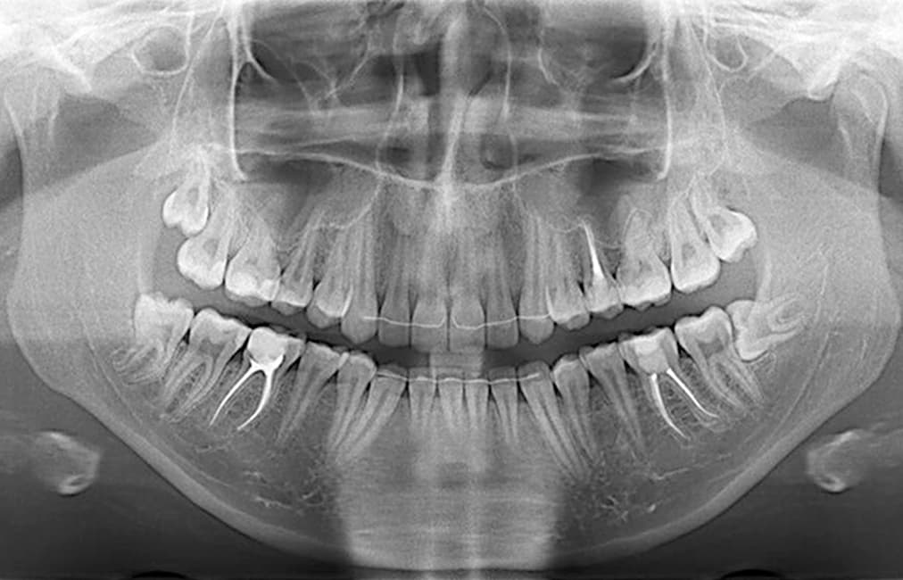
What does the orthopantomogram show?
Panoramic image of teeth shows the location of all dental roots and the condition of surrounding tissues, the quality of filling of canals, the presence of latent infection around root inflammations (granulomas, cysts), the degree of wisdom teeth eruption, the condition of jaw joints, reveals hidden carious cavities and diseases bone tissue.
- when planning a surgical operation;
- to assess the quality of therapeutic or surgical treatment;
- before installing dentures;
- before orthodontic treatment and in its process to control the correction of occlusion;
- with toothaches of unclear localization and a number of other symptoms
In which cases is a dental panoramic X-ray needed?
Orthopantomography is performed when planning dental treatment, especially if you need to treat several teeth and periodontal tissues, as well as:
Computed tomography (CT) of the teeth and jaw is one of the most reliable and informative research methods in modern dentistry. The tomography allows to take a picture in 3D, which allows dentists to diagnose and offer the patient the best solution accurately and quickly.
The new technology of cone-beam dental tomography is safe. The radiation is less than 30 μSv. This ultra-low dose is comparable to the natural background and is unable to cause any negative impact on the human body.